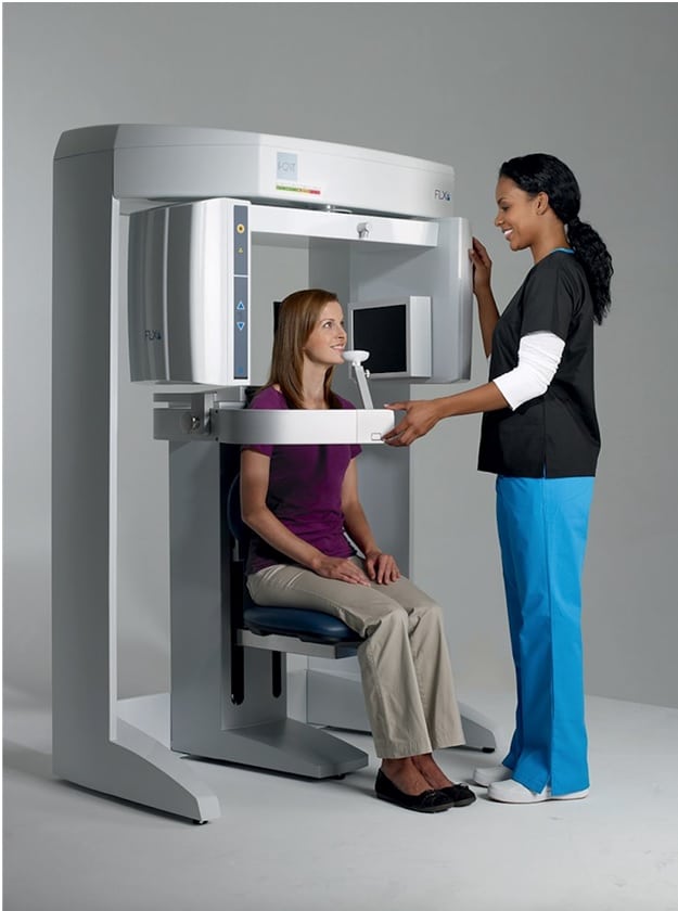
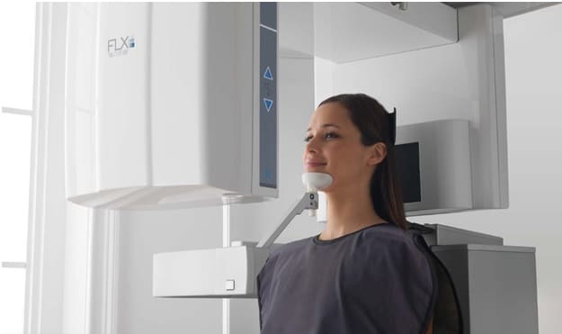
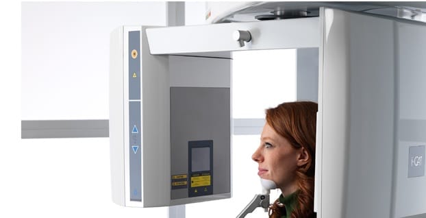
Every so often, dental professionals remark that the scans they’ve captured with their i-CAT CBCT machines do not exhibit high enough clarity or quality and question whether the machine needs to be calibrated or repaired to improve the image quality. It’s most likely, however, that many of these image quality issues are caused by improper patient positioning rather than equipment malfunction. These quick tips for patient positioning with an i-CAT cone beam system should help improve the quality of both your i-CAT CBCT and panoramic scans.

Positioning Patients for i–CAT CBCT Scans
First and foremost, patients must be positioned for minimal movement. Follow these steps to help prevent movement when capturing a dental cone beam scan:
– Utilize the head support, strap, and chin cup where possible.
– Use the footstool to stabilize the patient’s feet.
– Lower the chair elevation and tilt the chip up to eliminate concerns about shoulder clearance. You can perform a “Dry Run” to test for shoulder clearance.
- Adjust the chair height; set the elevation for the scan by aligning the horizontal laser light with the patient’s smile line.
- Close the gate.
- Ensure the patient’s chin is firmly seated in the chin cup.
- Secure the head support, with the head strap around the patient’s forehead.
- Adjust jaw tilt; the occlusal plane should be horizontal, the ala-tragus line should be horizontal.
Prior to taking the CBCT scan, instruct the patient:
– To remove all jewelry, including piercings.
– To remove glasses.
– Either to hold the bottom of the chair or to place the hands in their lap.
– Not to swallow during the scan.
– To breathe using shallow, even breaths.
– To keep their eyes closed.
– To remain as still as possible.
– To keep the teeth gently together.
– Note: Always use a lead apron during the dental ct scan.
Inform that patient that:
– The gantry will rotate around the head during the scan.
– The exposure buzzer will sound throughout the duration of the rotation.
Helpful tips:
– If the gate won’t close, try using the head support without the chin cup; keep the gate open and remove the chin support mechanism.
– Try using the chin cup without the head support.
– Slide the patient forward with the chair in one of the upper slots so that he or she can lean back into the scanner.
(For orthodontics, use only the head support so that the chin cup will not be visible.)
Scout Scans and Dry Runs
In order to verify correct patient positioning, you can perform a Scout Scan. Scout Scans expose the patient to a fraction of a second of radiation, but it is enough to provide an image that can be used to verify that the patient is positioned properly to capture a cone beam scan of the desired field of view.
A Dry Run does not administer any radiation to the patient, but it does demonstrate the motion of the machine and the rotation of the machine; it can be used to check for shoulder clearance.

Positioning Patients for a Panoramic X-ray Scan
Follow the steps below to ensure proper patient positioning when capturing a panoramic X-ray image with an i-CAT cone beam system:
- Place the chair in the upper slots; the back of the chair should not prevent the patient from sitting upright with an erect posture.
- If using the head support; loosen the locking knob and remove; Slide PAN head holder into place and tighten the locking knob.
- Insert the bite tip into the bit tip holder (narrow edge first). Turn the bite tip ¼ of a rotation to lock it into place.
- Insert the chin rest and bit tip holder into the positioning block.
- Have the patient sit; instruct the patient to sit with an erect, upright posture and to extend the neck up as straight as possible. Close the gate.
- Adjust the chair so that the patient’s chin is in the chin rest without affecting his or her upright posture.
- Tilt the patient’s head down until the occlusal plane is angled at 5 to 10 degrees to the horizontal laser.
- Ensure the patient is centered and facing forward with the Sagittal laser.
- Have the patient bite the groove in the bite tip and tell him or her to close the lips around it as though around a straw. (If necessary, adjust the bite tip).
- Close the head holder arms; temple pads should be adjacent to the patient’s temples. Adjust as necessary before tightening the knobs.
- Instruct the patient to swallow and hold with the tongue pressed against the roof of the mouth, holding the position until the exposure is completed.
Contact Renew Digital
Following these instructions, you should be able to capture high-quality, detailed images through i-CAT CBCT and panoramic scans. If you recently purchased an i-CAT system from Renew Digital and need additional assistance, please feel free to contact them for support.
If you are interested in purchasing an i-CAT X-ray system for your dental practice, such as the i-CAT FLX V17, i-CAT Next Generation, i-CAT FLX MV or i-CAT Precise, contact Renew Digital at 888-246-5611 or visit their website, RenewDigital.com. The leader in certified pre-owned digital dental systems, Renew Digital can save their customers anywhere from 30% to 50% off the price of new systems and include installation, training, and on-site support with every purchase.


